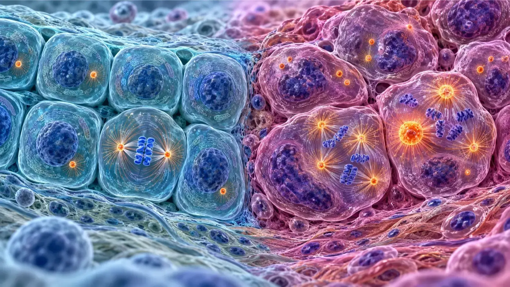
Medical accuracy note: CenSegNet has not cured cancer and is not currently a clinical diagnostic or treatment system. It is a research platform designed to analyze cellular structures in tissue images. Its findings could support future biomarkers and more personalized treatment strategies, but further validation and clinical research are required.
Artificial intelligence is rapidly changing how scientists investigate cancer. Instead of replacing oncologists, pathologists, or laboratory researchers, specialized AI systems can help them examine biological information at a scale and speed that would be extremely difficult to achieve manually. One promising example is CenSegNet AI, an open-source deep-learning platform developed by researchers at the University of Southampton in England.
CenSegNet—short for Centrosome Segmentation Network—was created to identify and measure centrosomes, tiny structures inside cells that help organize cell division. Abnormal centrosomes have been associated with cancer for more than a century, but examining them across large numbers of patient tissue samples has traditionally been slow and technically challenging.
That is where CenSegNet could make an important difference. In a 2026 study published in Nature Communications, researchers used the system to analyze 333,148 centrosomes across 911 breast-tissue sample cores from 127 patients. The AI distinguished different forms of centrosome abnormality and mapped how they were distributed in and around tumors. These patterns were associated with features such as tumor grade, hormone-receptor status, genetic alterations, lymph-node involvement, and overall survival.[1]
The findings do not mean that AI has discovered a cure for cancer. They show something more precise and scientifically responsible: AI may help researchers uncover cellular patterns that were previously too difficult to study at scale. In the future, this knowledge could contribute to improved biomarkers, more accurate risk assessment, and treatments selected according to the biological vulnerabilities of an individual tumor.
What Is CenSegNet AI?
CenSegNet is a modular computer-vision platform built to detect, outline, count, and measure centrosomes in complex tissue images. It can work with immunohistochemistry—the staining method routinely used in pathology laboratories—and immunofluorescence, an imaging technique that uses fluorescent labels to reveal specific cellular components.[1]
The platform combines several deep-learning methods. Its architecture uses YOLOv11 to locate likely centrosomes, U-Net to perform detailed pixel-level segmentation, and StarDist to identify individual cells and their boundaries. By combining these components, the system can connect centrosome measurements to particular cells and tissue regions.[1]
This is important because simply locating an object in an image is not enough for advanced cancer research. Scientists need to know its shape, size, number, cellular context, and location within a tumor. CenSegNet was designed to preserve this spatial information.
The software is also open source. The research team released the code and supporting resources publicly, allowing other scientists to test, inspect, and potentially extend the platform. Open access does not guarantee that a medical technology is clinically ready, but it can encourage independent validation and scientific collaboration.[1]
Why Centrosomes Matter in Cancer
Centrosomes are small cellular structures that act as major organizing centers for microtubules. Microtubules contribute to cell shape, movement, internal transport, and the formation of the mitotic spindle that separates chromosomes during cell division.[1]
Healthy cells normally control centrosome duplication carefully. A centrosome is copied once during the cell cycle so that chromosomes can be distributed correctly between two daughter cells. When this process is disrupted, a cell may develop too many centrosomes, oversized centrosomes, or other structural defects.
These abnormalities can contribute to errors in chromosome separation. The resulting genetic instability is closely connected to cancer biology. Centrosome abnormalities have been observed in breast, prostate, colon, ovarian, pancreatic, and other solid tumors, as well as in several blood cancers.[1]
Researchers generally discuss two broad categories:
- Numerical centrosome abnormalities: A cell contains an abnormal number of centrosomes.
- Structural centrosome abnormalities: Centrosomes become unusually large or display altered organization.
Before the CenSegNet study, these defects were often grouped together under one general concept. The Southampton researchers found evidence that numerical and structural abnormalities can behave as biologically distinct phenomena. Recognizing that difference could help scientists develop more informative biomarkers and better understand how tumors evolve.[1]
Why Studying Centrosomes Has Been So Difficult
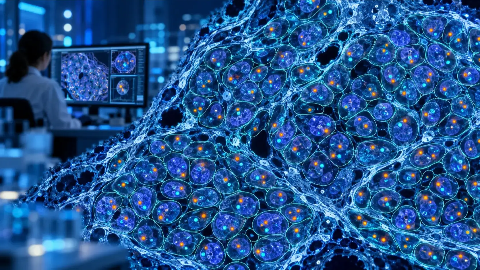
Centrosomes are extremely small. They must be highlighted with specialized laboratory stains and examined through microscopy. In a large tissue sample, scientists may need to identify thousands of these structures while also determining which cells contain them and whether they lie in normal tissue, at a tumor boundary, or deep within a tumor.
Manual annotation is time-consuming and can be affected by differences between observers. Earlier automated approaches helped with parts of the process, but many required manual correction, handled only one imaging method, or could not reliably distinguish the relevant cellular structures at single-cell resolution.[1]
CenSegNet was developed to address these limitations through automated, high-throughput analysis. The platform does not merely label an entire region as abnormal. It creates pixel-level outlines of centrosomes, associates them with cells, and preserves their positions within the tissue.
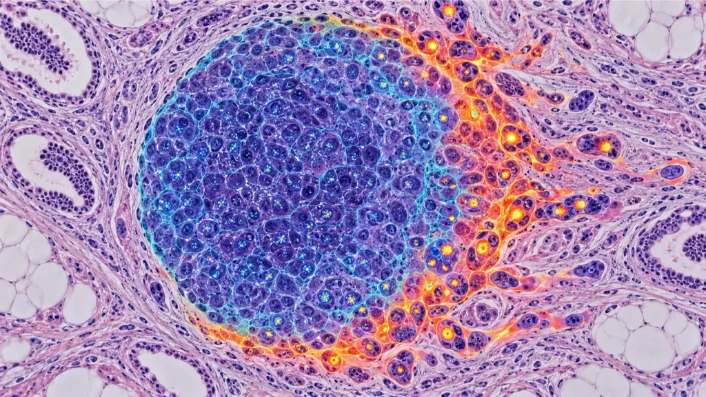
This spatial context matters because a tumor is not a uniform mass. Cells at its center can differ from those at its invasive edge, where cancer encounters and remodels surrounding tissue. An average measurement across the whole tumor could conceal clinically important differences.
How the University of Southampton Study Was Conducted
The study was led by researchers associated with the University of Southampton and University Hospital Southampton. The team constructed tissue microarrays containing 911 cores taken from tumors, tumor edges, adjacent regions, and normal breast tissue from 127 patients in the BeGIN breast-cancer cohort.[1]
To create training data, experts manually annotated 14,679 centrosomes in selected immunohistochemistry images. The researchers also built an immunofluorescence training set containing 1,285 annotated centrosomes from human mammary epithelial cells and mouse mammary tissue.[1]
CenSegNet was then compared with established segmentation architectures, including U-Net, SegNet, and DeepLabv3+. On combined test data, CenSegNet achieved a mean F1 score of 0.82, compared with 0.72 for U-Net, 0.68 for SegNet, and 0.65 for DeepLabv3+. Its predicted centrosome counts also showed very strong agreement with manually annotated reference data.[1]
Performance numbers should always be interpreted in the context of the tested datasets, but these comparisons suggest that CenSegNet handled the small size and visual complexity of centrosomes more effectively than the alternative models evaluated in the study.
Using the trained platform, the researchers automatically segmented 333,148 centrosomes. That scale allowed them to compare centrosome characteristics across tissue regions and connect them with clinical and molecular data.[1]
CenSegNet Revealed Two Different Patterns of Abnormality
One of the study’s most important results was that numerical and structural centrosome abnormalities did not simply rise and fall together. The researchers described them as mechanistically uncoupled, meaning they may represent separate dimensions of centrosome dysfunction.[1]
Their spatial distributions were also different. Numerical abnormalities—cells containing too many centrosomes—were more prominent in proliferative tumor cores. Structural abnormalities, including enlarged centrosomes, accumulated more strongly at invasive tumor margins.[1]
This discovery matters because the tumor core and edge experience different biological pressures. Cells at the edge interact with connective tissue, immune cells, blood vessels, and other features of the tumor microenvironment. Structural centrosome changes in this location might be related to invasion, movement, or adaptation to surrounding tissue. The study identified associations, but additional experiments will be needed to establish exactly how these relationships operate.
The findings challenge the idea that every centrosome abnormality carries the same meaning. A tumor characterized mainly by increased centrosome number may behave differently from one characterized by enlarged centrosomes. If future research confirms these differences, clinicians might eventually use these patterns to divide patients into more biologically meaningful groups.
Connections to Tumor Aggressiveness and Patient Outcomes
The study linked centrosome patterns with several established clinical features. Structural centrosome abnormalities were associated with higher tumor grade and lymph-node involvement. The researchers also found relationships involving hormone-receptor status, HER2 expression, and genomic alterations.[1]
Most notably, variation in structural centrosome abnormalities was associated with overall survival. The University of Southampton reported that patients whose tumor cores contained fewer enlarged centrosomes tended to have better overall survival.[2]
This result does not make CenSegNet a validated survival calculator. The cohort included 127 patients, and an association found in one retrospective dataset must be confirmed independently before it can guide individual care. Factors such as treatment history, tumor subtype, age, and other clinical characteristics can also influence outcomes.
Nevertheless, the association provides a strong reason to continue investigating centrosome-based biomarkers. A useful biomarker could help doctors estimate risk more precisely, identify aggressive disease, or choose between treatment strategies. CenSegNet gives researchers a practical way to measure the underlying cellular features consistently and at scale.
Could CenSegNet Help Cure Cancer?
The most accurate answer is that CenSegNet is helping cancer research, but it is not a cancer cure.
Cancer is not one disease with one biological cause. It includes many diseases driven by different combinations of genetic changes, cell types, environmental influences, and interactions with the immune system. Even within one person’s tumor, separate regions can contain distinct cell populations. That complexity is one reason why no single AI model should be presented as a universal cure.
CenSegNet could contribute to progress in several ways:
1. Discovering new biomarkers
If particular centrosome patterns consistently predict aggressive disease, treatment response, recurrence, or survival, those patterns may become useful biomarkers. CenSegNet enables researchers to test such hypotheses across large image collections.
2. Improving patient stratification
Patients with the same conventional diagnosis may have tumors with different cellular vulnerabilities. Centrosome phenotyping could add another layer of information to existing pathology, genomic testing, and receptor analysis.
3. Supporting precision oncology
Proteins such as PLK4, PLK1, AURKA, and components of the pericentriolar material influence centrosome number or structure. Some experimental cancer treatments target pathways connected to centrosome regulation. A future biomarker could help identify tumors most likely to respond to a particular centrosome-targeting strategy.[1][2]
4. Accelerating laboratory research
Automated analysis can allow scientists to evaluate far more cells than would be realistic through manual annotation alone. This may shorten the process of testing hypotheses and comparing experimental treatments.
5. Revealing intratumoral heterogeneity
By mapping abnormalities in the center and at the edge of tumors, researchers can study why some regions invade, resist treatment, or evolve differently. Understanding this heterogeneity could eventually inform combination therapies.
These contributions are meaningful steps toward better cancer care. However, there is a long scientific path between discovering a pattern and proving that using it improves patient outcomes.
How CenSegNet Could Advance Personalized Cancer Treatment
Personalized cancer treatment aims to match therapy to the characteristics of a patient and tumor. Breast-cancer care already uses information such as estrogen-receptor status, progesterone-receptor status, HER2 expression, tumor grade, stage, and selected genomic tests.
CenSegNet could potentially complement these tools by adding a spatial map of centrosome biology. For example, two tumors may have similar receptor classifications but very different patterns of structural abnormality at their invasive edges. If those patterns are eventually shown to predict metastasis or sensitivity to a drug, they could help refine treatment decisions.
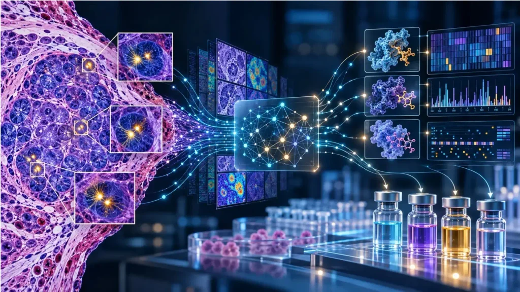
The Southampton team plans to combine CenSegNet measurements with genomic, transcriptomic, and proteomic information. Genomics examines DNA, transcriptomics studies RNA activity, and proteomics measures proteins. Integrating these data could reveal why a visible centrosome pattern appears and which molecular pathway sustains it.[2]
This type of multimodal analysis is an important direction for medical AI. Images show what tissue looks like, while molecular data can provide clues about why it behaves that way. AI may help researchers connect those layers, but prospective studies must still demonstrate clinical value.
CenSegNet May Extend Beyond Breast Cancer
The primary clinical analysis focused on breast-cancer samples, but the researchers also tested the platform in human kidney, colon, and appendix tissues. The results indicated that the framework can generalize to different epithelial tissues and imaging contexts.[1]
That flexibility creates opportunities beyond a single disease. Centrosome abnormalities are found in numerous cancers, and organelle-level analysis could also be relevant to noncancerous epithelial diseases. Still, performance in a new tissue cannot be assumed. Each application will require suitable staining, reference annotations, validation datasets, and expert review.
Because CenSegNet is modular, researchers may be able to adapt or expand it as imaging technologies and deep-learning methods improve. Its value will depend not only on algorithmic accuracy but also on reproducibility across laboratories and patient populations.
Limitations and Questions That Still Need Answers
Exciting findings should be considered alongside their limitations.
First, CenSegNet is not ready for routine clinical use. The University of Southampton explicitly describes future diagnostic and treatment applications as possibilities rather than existing capabilities.[2]
Second, the major patient analysis involved a cohort of 127 people. That was sufficient to reveal promising associations, but larger and more diverse cohorts are needed to determine whether the findings generalize across hospitals, demographic groups, breast-cancer subtypes, staining protocols, and scanners.
Third, retrospective association is different from clinical utility. Even if a centrosome pattern correlates with survival, researchers must establish whether using that information changes treatment decisions and leads to better outcomes.
Fourth, tissue preparation can affect image appearance. Differences in staining quality, antibodies, microscopes, and laboratory procedures can reduce an AI system’s performance when it is moved to a new institution.
Fifth, human oversight remains essential. Pathologists and scientists must review outputs, investigate unusual cases, and interpret measurements alongside the complete clinical picture.
Finally, researchers must consider data governance, transparency, bias, and regulatory requirements before any AI-based pathology tool is deployed in healthcare. Open-source access supports transparency, but it does not replace formal clinical evaluation.
Why This Research Is Important
CenSegNet represents more than a faster counting tool. It demonstrates how AI can transform a difficult cellular observation into structured, spatially resolved data. By measuring hundreds of thousands of centrosomes, the system allowed researchers to see that different abnormalities occupy different tumor regions and carry different clinical associations.
This is a good example of the realistic role of AI in medicine. The technology does not independently diagnose a patient, invent a proven therapy, or eliminate the need for scientists. Instead, it expands what researchers can observe and quantify. That improved visibility can generate better questions, reveal possible biomarkers, and identify biological vulnerabilities worth targeting.
The distinction is important. Sensational claims about an “AI cancer cure” may attract attention, but they can mislead readers and weaken trust. The genuine achievement is already impressive: CenSegNet offers a scalable method for studying a long-recognized hallmark of cancer at a level of detail that was previously difficult to achieve.
The Future of CenSegNet AI in Cancer Research
The next stage will require independent validation and collaboration among cell biologists, oncologists, pathologists, data scientists, and clinical-trial experts. Researchers will need to test whether centrosome patterns remain informative in larger cohorts and whether they predict response to specific therapies.
Another priority will be combining centrosome maps with genetic and protein information. This could reveal therapeutic targets or help explain why some tumors develop numerical abnormalities while others develop structural ones. Researchers may also investigate whether changes in centrosome patterns over time reflect treatment resistance or disease progression.
If future studies are successful, CenSegNet-derived measurements might become part of a broader decision-support system. A pathologist could potentially combine conventional tissue features, molecular test results, and AI-generated spatial information to build a more complete tumor profile.
That possibility remains a research goal, not a current clinical service. Yet the study provides a credible foundation. Its peer-reviewed publication, transparent methodology, large-scale cellular analysis, and open-source software make CenSegNet a noteworthy development in AI-assisted cancer research.
Final Thoughts
CenSegNet AI shows how deep learning can help scientists investigate cancer at an extraordinarily detailed level. Developed by University of Southampton researchers, the platform detects and measures centrosomes across complex tissue images and links their abnormalities to individual cells and tumor locations.
In breast-cancer samples, it revealed that having too many centrosomes and having enlarged centrosomes are not simply versions of the same problem. These abnormalities displayed different spatial patterns and different relationships with clinical features. Structural abnormalities were associated with aggressive disease characteristics and overall survival, creating promising directions for biomarker and precision-oncology research.[1]
The technology should not be called a cure for cancer. It is a research instrument that may help scientists understand tumors, discover biomarkers, and eventually select treatments more intelligently. That is a more careful claim—but also a powerful one. By revealing patterns that human researchers could not feasibly map at the same scale, CenSegNet may help move cancer science toward more precise and personalized care.
Frequently Asked Questions
What does CenSegNet mean?
CenSegNet stands for Centrosome Segmentation Network. It is a deep-learning framework that detects and measures centrosomes and maps them within cells and tissues.[1]
Did CenSegNet cure cancer?
No. CenSegNet has not cured cancer. It is a research tool that helps scientists analyze cellular structures associated with cancer. Its discoveries may contribute to future biomarkers and personalized treatment strategies.
Who created CenSegNet AI?
CenSegNet was developed by a multidisciplinary research team led from the University of Southampton, working with University Hospital Southampton and other collaborators. Dr. Salah Elias led the research.[1][2]
What type of cancer did the researchers study?
The main patient analysis examined breast cancer. The researchers analyzed 911 tissue cores from 127 patients and automatically segmented more than 333,000 centrosomes. They also demonstrated the platform in kidney, colon, and appendix tissues.[1]
Is CenSegNet being used in hospitals?
It is not currently ready for routine clinical use. The platform must undergo further independent and clinical validation before its measurements can guide patient care.[2]
How could CenSegNet support personalized medicine?
It could help researchers identify centrosome patterns connected to tumor aggressiveness, prognosis, or drug sensitivity. If validated, such patterns might complement existing pathology and molecular tests when selecting treatments.
Is CenSegNet open source?
Yes. The researchers released CenSegNet as an open-source Python resource, supporting scientific evaluation and further development.[1]
References
[1] Primary research article: Cheng, J., Fan, K., Bailey, M., et al. “CenSegNet: a generalist high-throughput deep learning framework for centrosome phenotyping at spatial and single-cell resolution in heterogeneous tissues.” Nature Communications, volume 17, article 8713 (2026). Published July 15, 2026. https://doi.org/10.1038/s41467-026-75393-y
[2] Primary institutional research release: University of Southampton. “AI reveals hidden patterns inside breast cancer.” Published August 18, 2026. https://www.southampton.ac.uk/news/2026/08/ai-reveals-hidden-patterns-inside-breast-cancer.page
[3] Primary software repository: SKELab, University of Southampton research team. “CenSegNet.” GitHub repository linked by the peer-reviewed study. https://github.com/SKELab/CenSegNet/
Editorial disclaimer: This article is for educational and informational purposes only. It does not provide medical advice, diagnosis, or treatment. Anyone with questions about cancer screening or treatment should consult a qualified healthcare professional.